IBL now offers Full Production — design, print & sterilize in one place. See our services →
System Applications Process Submit case
← All solutions
Human Maxillofacial Reconstruction

Built to the exact contour of the bone.

IBL designs and fabricates patient-specific titanium subperiosteal frameworks for severe ridge atrophy, post-oncologic defects, and failed endosteal implant salvage.

0.3mm
Mesh-to-bone tolerance
9–12d
Design turnaround
4–8
Fixation points, per case
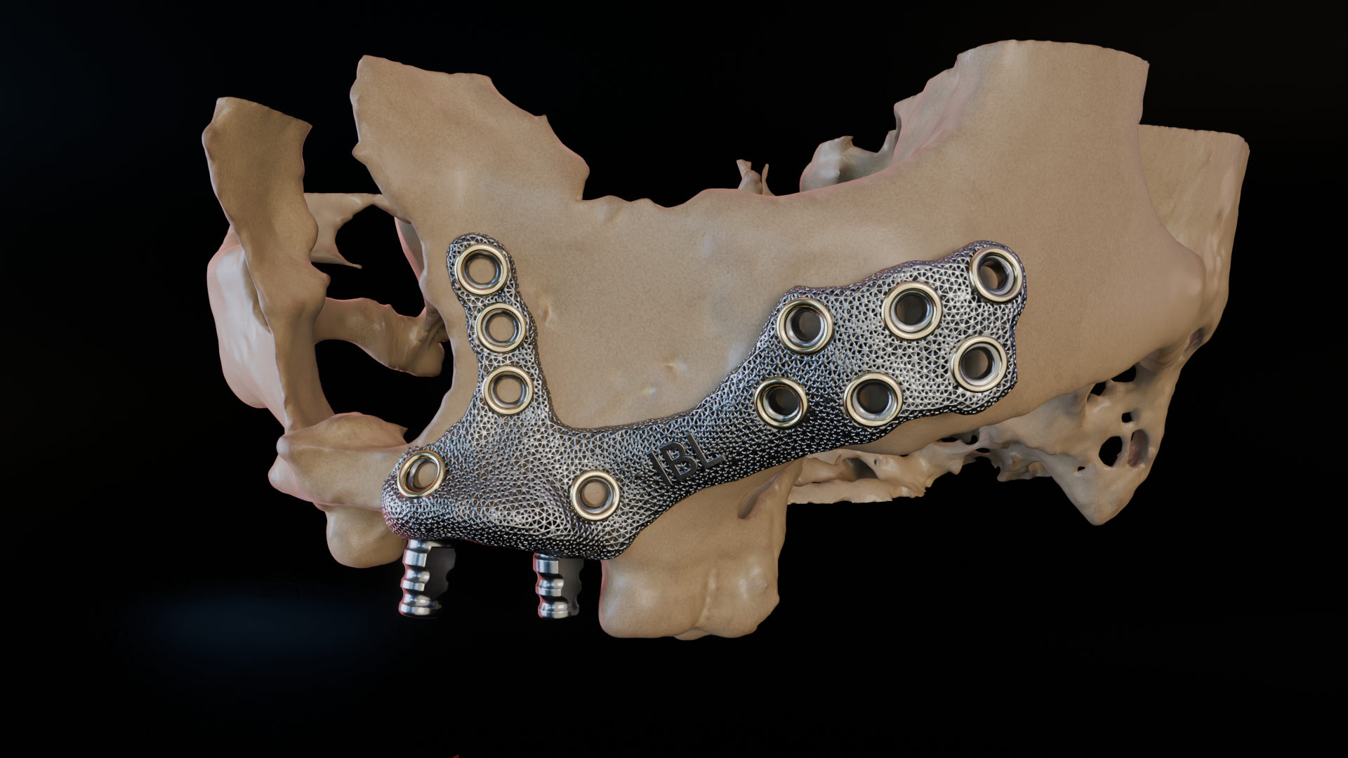
Titanium subperiosteal framework for both upper and lower jaw arches, case model
CASE MODEL — MAXILLA / MANDIBLE, DUAL ARCH
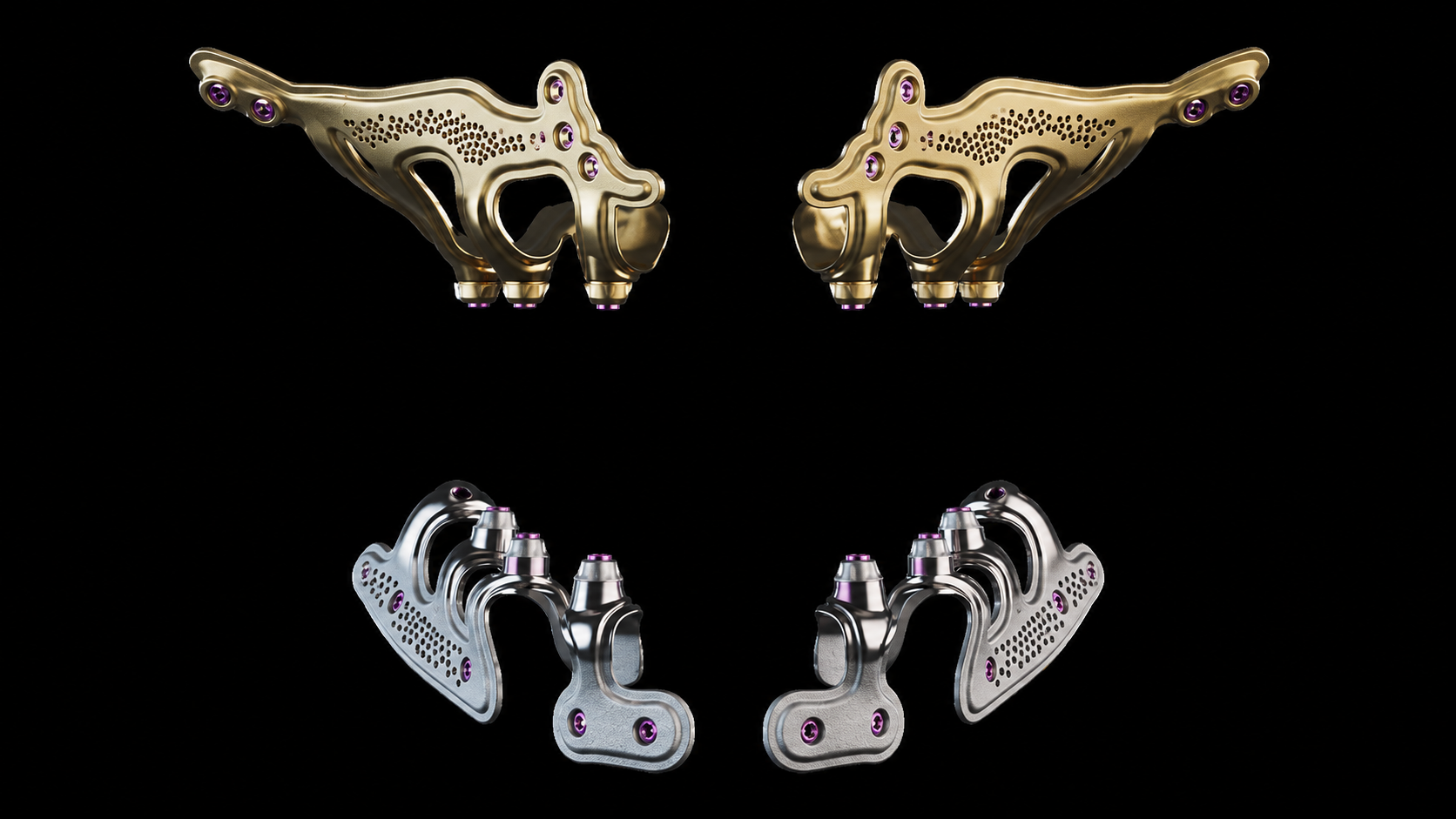
Maxillofacial reconstruction

Engineered for the human maxilla and mandible

For patients with severe ridge resorption or bone loss where conventional endosteal implants can't get adequate purchase, we design a mesh framework that rests on the bone surface beneath the periosteum, distributing load across the residual ridge instead of relying on it for anchorage.

  • IndicationSevere alveolar atrophy, post-oncologic defects, failed endosteal cases
  • MaterialGrade 23 titanium mesh, laser-sintered
  • Design inputCone-beam CT, 0.2mm slice thickness
  • Surgeon fitReviewed digitally before fabrication, no chairside adjustment
Case Reference — Mandibular
Framework weight4.8–6.2 g
Strut thickness0.6 mm
Fixation points4–8, per case
Design turnaround9–12 business days
SterilizationSteam, pre-packaged

Applications

01 / INDEX
Case typeFrequencyNotes
Severe ridge / bone atrophyPrimary indicationMost common referral pathway
Post-tumor resection defectSupportedCoordinated with oncologic surgery timeline
Failed endosteal implant salvageCommonWhere residual bone can't support new fixtures
Traumatic fracture non-unionOccasionalCase-by-case review
Congenital / developmental defectOccasionalCase-by-case review

From scan to placement

02 / PROCESS
01

Imaging

Referring surgeon submits a CT scan of the affected bone, along with case history and desired outcome.

CT / 0.2mm slices
02

Digital design

Our engineers model the bone surface in CAD and design a mesh framework that follows its exact topography, then route fixation points around anatomical structures to avoid.

CAD / surface mesh
03

Surgeon review

The design is sent back for digital sign-off before fabrication begins — this is the point to request changes to coverage, strut pattern, or fixation layout.

Digital proof
04

Fabrication

The approved design is laser-sintered from titanium, finished, and sterile-packaged.

Ti-6Al-4V ELI
05

Placement

The framework is seated directly on the bone surface beneath the periosteum and secured at the pre-planned fixation points — no intraoperative shaping needed.

Subperiosteal

Subperiosteal vs. endosteal

03 / COMPARISON
FactorSubperiosteal (IBL)Endosteal
Bone volume requiredMinimal — rests on bone surfaceSufficient height/width needed
FitPatient-specific, designed pre-opStandard sizes, shaped chairside
Best suited forSevere atrophy, salvage, irregular defectsAdequate residual bone
PlacementSingle surgical stageOften staged, with healing interval
Submit a case

Send us a CT scan.

Upload the scan and your contact details, and our design team will follow up with a feasibility review and timeline — usually within 2 business days. Choose Digital design only if you'll fabricate in your own lab, or Full production if you want a finished implant. See both services →

Clinical inquiriescases@ibl-lab.example
Phone+1 (555) 019-2244
HoursMon–Fri, 8:00–18:00
Accepted filesDICOM (.dcm), zipped CT folder (.zip), STL — up to 200MB
Have a veterinary case instead? Visit our veterinary site →

Choose file — .dcm, .zip, or .stl

Files are transferred securely and reviewed only by our design team.